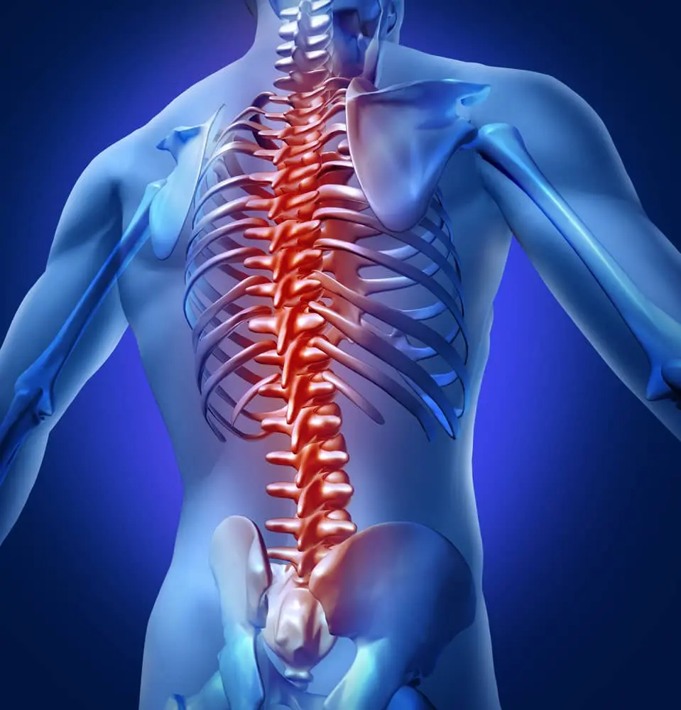
Spinal Canal Narrowing
(Spinal Canal Stenosis)
On this page, we will explain the details of spinal canal narrowing, its differences across spinal canal regions (cervical spinal canal narrowing, lumbar spinal canal narrowing, and thoracic spinal canal narrowing), and narrow canal surgery. We wish you all healthy days.
What Is Spinal Canal Stenosis in the Lower Back, Mid-Back, and Neck?
The spinal cord runs within a bony canal formed by the vertebra. This spinal canal extends downward starting from the base of the skull. It is divided into four regions: cervical (neck), thoracic (mid-back), lumbar (lower back), and sacral. There are 7 vertebra in the cervical region, 12 in the thoracic region, and 5 in the lumbar region. Other important anatomical structures include the intervertebral discs between the vertebra, the facet joints that allow the vertebra to articulate with each other, and the strong ligaments and muscle layers located behind the vertebral bodies.
Spinal canal narrowing (spinal stenosis) occurs when the canal behind the vertebra becomes constricted and compresses the spinal cord as it travels from top to bottom, as well as the nerve roots exiting it at different levels. This is a degenerative process that progresses with age. All the anatomical structures mentioned above contribute to this degeneration. Over time, patients may develop narrowing of the spinal canal in the cervical, thoracic, or lumbar regions.
With aging, the intervertebral discs lose water and height. In overweight patients, the facet joints enlarge inward to bear the increased load. The ligaments behind the spine gradually thicken and may calcify, and the ligament in front of the spinal cord can also calcify. In addition, the ligamentum flavum (a yellow ligament at the back of the spinal canal) thickens with age, compressing the spinal cord from behind. All these changes contribute to spinal canal narrowing.
Symptoms of Spinal Canal Narrowing in the Lower Back, Mid-Back, and Neck
Because lumbar spinal canal narrowing usually develops slowly, it may not cause symptoms at first. However, as the disease progresses, it can significantly reduce quality of life and limit daily activities. When symptoms become obvious, the canal diameter is often already severely narrowed and the spinal cord and nerve roots are firmly compressed.
Patients may experience low back or mid-back pain, weakness, and numbness in the legs or feet. One hallmark sign is cramping or tightening in the legs after walking a certain distance. Over time, the distance a patient can walk decreases, and in advanced stages, even walking around the house can become difficult. These symptoms, which appear with walking, partially improve after a period of rest, but reappear after walking the same distance again.
In addition, patients may experience pain starting in the hip and radiating down the leg and into the foot, like sciatica from a herniated disc. Many patients with spinal canal narrowing also have difficulty lying flat on their backs. In later stages, patients tend to walk leaning forward. Bending forward slightly increases the diameter of the spinal canal and may provide temporary relief.
In cervical spinal canal narrowing, symptoms may include numbness, pain, and tingling in the hands or arms, difficulty performing fine motor tasks, gait disturbance, and in more advanced stages, urinary or faecal incontinence due to impaired bladder and bowel control.
How Is Spinal Canal Narrowing Diagnosed?
Various imaging methods are used to diagnose spinal canal narrowing. One of the first is plain radiography (X-ray). On X-rays, we can evaluate the alignment of the vertebra, their anatomical structures, the diameter of the foramina where the nerve roots exit, degenerative changes in the spine, any slippage (spondylolisthesis) of the vertebra, and their anatomical relationships with each other.
Three-dimensional computed tomography (CT) provides more detailed information about these anatomical structures. 3D images allow us to visualize the internal structure of the spinal canal more clearly. With CT, it is also possible to take precise measurements in advance to decide on the size of screws, rods, and other fixation devices that may be needed during surgery.
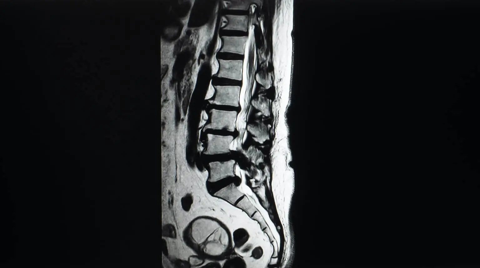
Magnetic resonance imaging (MRI) has become the gold standard diagnostic method in recent years. MRI allows us to evaluate the intervertebral discs, the facet joints, the ligaments that hold the vertebra together, the dural sac surrounding the spinal cord, and the nerve roots exiting from it. A spinal canal diameter that falls below a certain threshold on MRI is considered a sign of disease progression.
Electrophysiological studies such as electromyography (EMG) and somatosensory evoked potentials (SSEP), commonly referred to as “nerve conduction tests,” can be used to evaluate how well the spinal cord conducts signals and to determine which nerve roots are under pressure and how severely they are affected. These tests also help differentiate spinal canal narrowing from other diseases that can affect the spinal cord and nerves.
What Is Done During Spinal Canal Narrowing Surgery?
In spinal canal decompression surgery, the laminae — the bony plates forming the back part of the vertebra on both sides — and the yellow ligament (ligamentum flavum) beneath them are removed to widen the spinal canal. To maintain spinal stability and prevent future slippage, spinal fusion (fixation of the vertebra in the operated segment) with implants may be performed during the same procedure.
In patients with moderate spinal canal narrowing, instead of removing both laminae, surgery may be performed through a unilateral (one-sided) approach. Using microsurgical techniques, the surgeon removes the lamina on one side but decompresses the spinal canal on both sides, fully relieving the compression.
Because spinal canal narrowing is a highly degenerative process, some patients develop vertebral slippage (spondylolisthesis). In such cases, the bone behind the spine is removed on both sides to widen the canal, and then the affected vertebra are stabilized with implants (screws and rods).
In cervical spinal canal narrowing, the problem can be addressed with either anterior (front) or posterior (back) surgical approaches. The choice of approach is made after neurological examination and review of imaging and electrophysiological studies.
If the main problem is a disc pressing on the spinal cord in the neck region, surgery may be limited to removing only the affected disc, without interfering with the two neighboring vertebra. Sometimes, however, there is significant calcification of a strong ligament located behind the vertebral bodies that compresses the spinal cord from the front. In that case, the affected vertebral body and the disc tissue are removed together (corpectomy). They are replaced with a bone graft or a cage-like prosthesis that restores the height and function of the removed vertebral segment. A titanium plate is then placed in front to hold everything in place and prevent movement or dislodgement.
Treatment of Spinal Canal Narrowing in the Lower Back, Mid-Back, and Neck
For patients in the early stages of the disease, non-surgical treatment is usually recommended first. These options include rest, medications, injections into or around the spinal canal, and physical therapy.
Medication may range from simple pain relievers to stronger narcotic pain medications in more severe cases. The appropriate drug and dosage must be determined by a physician.
Epidural and transforaminal injections performed by neurosurgeons, anaesthesiologists, or pain specialists (algologists) are also non-surgical alternatives. In these procedures, a mixture of corticosteroid (“cortisone”) and local anaesthetic is injected either into the epidural space (the space outside the membrane that surrounds the spinal cord and nerves) or into the nerve root canal where the nerve exits the spine to travel down the leg.
The main goals of physical therapy, prescribed and supervised by a physical medicine and rehabilitation specialist, are to relieve or significantly reduce pain, strengthen the supporting muscles, and increase the patient’s mobility and functional independence.
In patients with neurogenic claudication — where walking distance is severely limited due to cramps, tightening, and weakness in the legs or feet — quality of life can be greatly reduced. Sometimes, more serious problems such as urinary or fecal incontinence may develop.
Thanks to advances in technology and the increased use of microsurgical techniques, spinal stenosis surgery can now be performed more safely and comfortably, with better outcomes. The primary goal of surgical treatment is to decompress the spinal cord and nerves in the neck, thoracic, or lumbar regions. This procedure is called decompression. If there is instability or a risk of instability, fusion surgery (stabilization with screws and rods) may be added.
Recovery After Spinal Canal Narrowing Surgery
After cervical, thoracic, or lumbar spinal stenosis surgery, patients are generally mobilized (helped to stand and walk) about 6 hours after the operation. Hospital stay is typically 2 to 4 days. There is usually no problem with traveling home by car in a seated position after discharge.
Patients can resume daily activities while wearing the prescribed brace or corset. They should pay attention to proper body mechanics, particularly when sitting down and standing up.
In the first days after surgery, pain, burning, or stinging at the surgical site are common and not a cause for concern. These symptoms can usually be controlled with pain medications prescribed after the operation and short periods of rest.
Patients should ensure that their mattress is suitable for spinal health. After discharge, they should avoid sleeping on unsuitable surfaces such as couches or armchairs.
When getting into bed, patients should first sit on the edge of the bed with support, then, using their hands, lower their head onto the pillow and gently roll onto their side before turning into a supine (on the back) position. They may lie on their back or side as tolerated.
When getting out of bed, they should first roll onto their side, then move into a sitting position using their hands for support and finally stand-up using support from their knees.
Generally, medications given at discharge should not be continued or restarted later unless specifically instructed by the physician.
Patients who have undergone spinal stenosis surgery at any level should use a Western-style (raised) toilet rather than squatting toilets. They should put on their shoes while sitting. Women should avoid very high heels or completely flat shoes; medium-height, cushioned heels are more appropriate.
Patients who have had lumbar or thoracic spinal stenosis surgery should use a step or an appropriate platform when reaching for objects at a height, rather than stretching or climbing insecurely. They should not drop into chairs or armchairs; instead, they should sit down slowly, using their hands or knees for support. When standing up, they should push up using their knees or the edges of the seat.
Patients who have had thoracic or lumbar surgery can start walking short distances after about 7 days. Initially, walking sessions should last 20–30 minutes. Over time, this can be increased by 5–10 minutes per day, depending on tolerance and medical advice.
Patients who did not have screws (implants) placed during surgery and who work in an office setting can usually return to work after about 20 days. Those who perform heavy physical labour can usually return after about 60 days.
In the first 45 days after surgery, patients should avoid lifting heavy objects. After that, they should not carry more than about 5 kg in total (distributed between both hands). When lifting any weight, they should squat down, keep the object close to the body, and use the leg muscles instead of bending at the waist.
Postoperatively, patients should avoid weight gain and, if they are overweight, should aim to lose excess weight. Getting help from a dietitian is recommended.
The most suitable forms of exercise after spinal surgery are walking and, where possible, swimming.
Patients should not drive for the first 20 days after surgery. After that, they may drive short distances within the city, provided their doctor approves. Long-distance driving (for example, intercity travel) is usually considered safe about 30 days after surgery.
During long car journeys, it is important for spinal health to stop approximately every 1.5 hours, get out of the car, take a 10-minute break, walk a little, and then continue.
Patients can usually take short flights about 7 days after surgery. Longer flights — such as to Europe or transatlantic destinations — are generally allowed after about 30 days, during which time they should stand up and walk around the cabin every 1.5 hours.
Patients who have undergone spinal canal narrowing surgery at any level should use an office chair with good lumbar (lower back) support and stand up at least once every hour to walk around for 5–10 minutes.
Patients who have had thoracic or lumbar spinal stenosis surgery should avoid contact sports or activities with a high risk of falls or collisions.
In addition, patients who have undergone cervical spinal stenosis surgery should use a neck collar for about 10 days. Office workers can usually return to work on day 10, whereas those with physically demanding jobs should rest for approximately 4 weeks.
Patients should not carry more than 3 kg in their hands during the first 4 weeks after cervical surgery. Within the first year, they may gradually increase to carrying up to 7 kg total, divided between both hands, as advised by their doctor.
Driving should be avoided for 10 days after cervical spinal surgery. Short flights are allowed shortly after surgery, while longer flights (to Europe or overseas) are usually recommended only after about 30 days.
Patients who have undergone cervical spinal stenosis surgery should avoid contact sports for the first 4 months. Walking is recommended as the primary form of exercise during the first 20 days, after which they may begin swimming for fitness, if approved by their surgeon.
After cervical spinal surgery, patients should adjust their computer screens so that the top of the screen is at or slightly below eye level.
Patients who have undergone cervical, thoracic, or lumbar spinal stenosis surgery can start the prescribed exercise program approximately 60 days after the operation. Mild pain during these exercises is common at first but should gradually decrease and disappear over time.
In the postoperative period, patients should protect their spinal health and avoid activities that provoke pain in the neck, mid-back, or lower back. Factors that positively influence spinal health after surgery include performing the recommended exercise program regularly and avoiding weight gain.

