Spinal Canal Narrowing
On this page, we will try to explain the details of spinal canal narrowing, its differences according to the spinal canal regions (cervical spinal canal narrowing, spinal canal narrowing in the back and spinal canal narrowing in the waist) and narrow canal surgery.I wish you all healthy days.
What is Spinal Canal Narrowing in the Waist, Back and Neck?
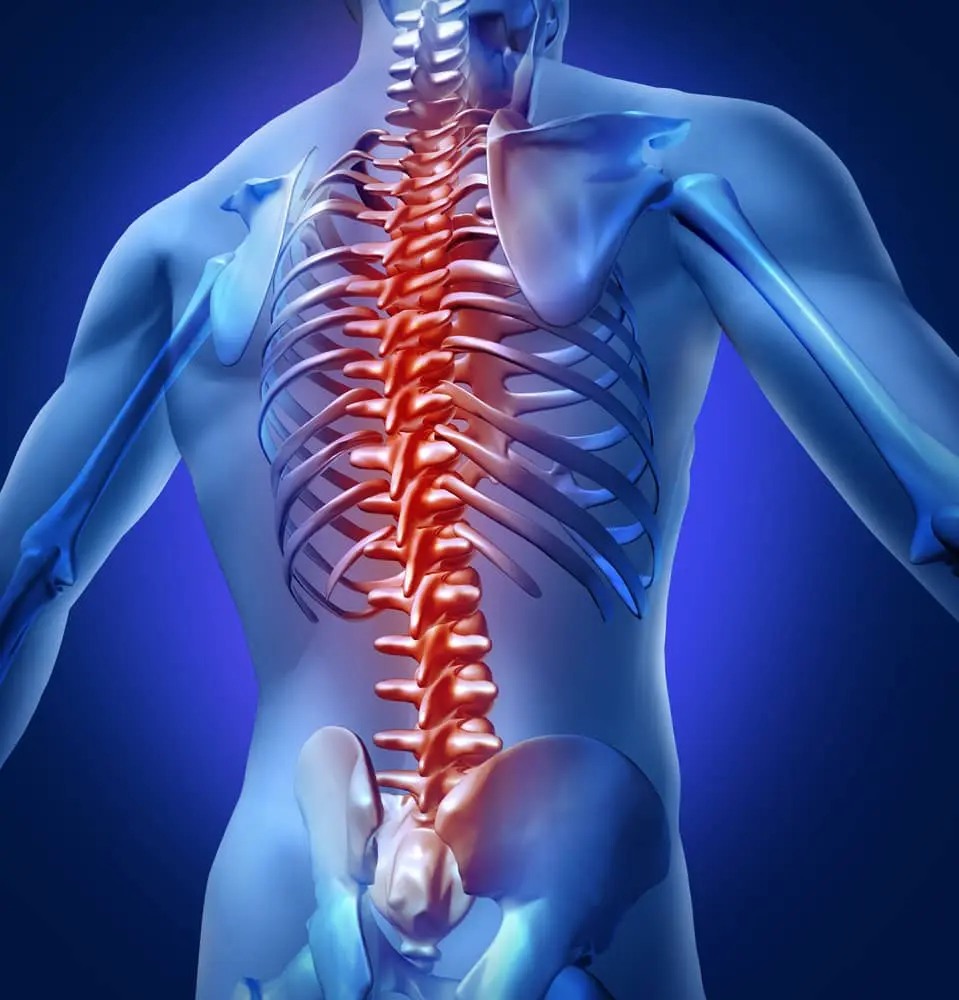
The spinal cord is located in the canal behind the structures called the spine. The spinal canal extends downwards, starting from the lower part of the head. There are 4 separate regions in the spinal canal: neck, chest, waist and sacral. There are 7 vertebrae in the neck area, 12 in the back area and 5 in the waist region. Other anatomical formations in the vertebrae are the structures we call discs between the vertebrae, the facet joints that allow the vertebrae to articulate with each other, and the strong connective tissues and muscle layer behind the body of the spines.
Spinal canal narrowing occurs when the canal behind the vertebrae narrows and compresses the spinal cord, which moves from top to bottom, at various levels and with the pressure it makes to the nerve roots. This process is a degenerative process that increases with age.All the anatomical structures mentioned above contribute to this degenerative process, and patients after a while are faced with the problem of narrowing of the spinal canal of the neck, back or lumbar spinal cord.
The height of the discs between the vertebrae decreases by losing water as they age, in overweight patients, the facet joints grow inward to carry the excess load, the connective tissue behind the spine calcifies over time and the spinal cord is calcified from the front, and the yellow connective tissue behind the spinal canal thickens with increasing age, compressing the spinal cord from the back and causing spinal canal narrowing.
Spinal Cord Canal Narrowing Symptoms in the Waist, Back and Neck
Since the narrowing of the lumbar spinal canal is a slowly developing process, it may not cause complaints and findings at first. However, with the progression of the disease over time, deterioration in quality of life and significant restriction in daily activities may occur. When these findings occur in patients, the spinal canal diameter is often severely narrowed and the spinal cord and the nerve roots emerging from it are stuck. Back and lumbar pain, loss of strength, and numbness in the legs or feet may occur. One of the important signs of the disease is the cramps and contractions that occur in the legs of the patients after walking a certain distance. Over time, the distance that patients walk decreases and they begin to have difficulty walking even within the house in later periods. These symptoms, which occur with walking in patients, partially decrease with a certain period of rest, then the same symptoms reappear after they start walking again and travel a certain distance. In addition, patients may also have pain that starts from the hip and spreads to the leg and foot, just like in the herniated disc. Patients with spinal canal narrowing often also have trouble lying on their backs. In later periods, these patients tend to walk by leaning forward. Because the patient's leaning forward creates some expansion in the spinal canal, causing temporary relief of the patient. In cervical spinal canal narrowing, numbness, pain, tingling sensation in the hands or arms, problems in doing skillful tasks in the future, gait disorder, and in many later stages of the disease, urinary and fecal incontinence problems may occur as a result of deterioration in bladder and bowel functions.
How Is Spinal Cord Canal Narrowing Diagnosed?
Various imaging methods are used in the diagnosis of spinal canal narrowing.One of them is plain graphy or x-ray film, as it is popularly called.On plain X-ray, the arrangement of the lumbar spines, their anatomical structures, the diameter of the channels where the nerve roots exit, the degenerative changes in the spine and the slippage in the spines and the anatomical relationship of the vertebrae with each other are evaluated.In three-dimensional computed tomography, which is another diagnostic method, more detailed information about the anatomical structures mentioned above can be obtained.In addition, 3D images allow us to visually see the internal structure of the spinal canal in more detail.With this examination, the size of the systems for fixing the spine, such as the screws that should be used in the surgery and the rods connecting these screws, will be used by pre-measured measurements.
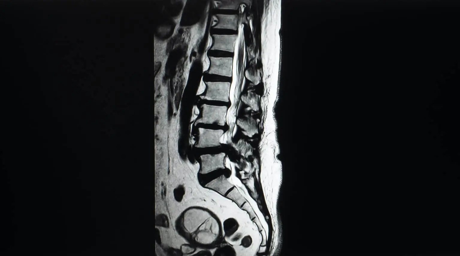
The diagnostic method, which has been seen as the gold standard in recent years, is magnetic resonance imaging.With this examination, it is possible to evaluate the disc structures located in the distance between the vertebrae, the regions where the vertebrae joint with each other and the ligament structures holding the vertebrae together, the spinal cord sac and the nerves coming out of it.In magnetic resonance imaging, the fact that the diameter of the spinal canal has fallen below a certain amount is considered as a sign that the disease is progressing.Among the people we call electrophysiological examination, electromyography and somatosensory stimulated potential examinations, which are called nerve measurement tests, can be used to evaluate the conduction of the spinal cord and which nerve coming out of the spinal cord is under pressure and to what extent it is affected by this pressure.With this method of examination, it is also possible to make a differential diagnosis with other diseases of the spinal cord.

What is done in Spinal Cord Canal Narrowing Surgery?
In spinal canal narrowing surgery, the bones called lamina on both sides, which form the posterior part of the spine, and the yellow connective tissue called ligamentum flavum just below it are taken and the spinal canal is expanded, and in order to protect the dynamics of the spine and to prevent spinal slippage in the future, the patients can be implanted and fusion, which is the fixation process of the spines in the operation area, can be performed.
In patients with more moderate spinal canal narrowing, instead of removing the bone tissue called lamina on both sides of the spine, the lamina on one side is removed by performing a surgical approach on one side.However, using microsurgical technique, spinal canal expansion surgery is performed on both sides and the compression here can be completely eliminated.Since spinal canal narrowing is a highly degenerative process, in some patients the spine may shift over each other and in this case, the bone tissue behind the spine is removed bilaterally and the spinal canal is widened and the vertebrae in the surgical site are fixed with an implant and surgery is performed.
In spinal canal narrowing surgeries in the neck area, it is possible to eliminate this problem with front or back surgeries.However, it is necessary to decide this after neurological examination and imaging and electrophysiological tests.If only disc structures that press on the spinal cord in the neck area are performed, surgical intervention is performed only in the disc section without any intervention for the two vertebrae adjacent to that disc.Sometimes there may also be calcification of a very strong ligament extending behind the spine, which puts pressure on the spinal cord from the front.In this case, the affected spinal body and disc tissue are removed together.In its place, structures called grafts consisting of bone tissue or cage-like prostheses that will replace the spine are placed.Then, titanium plaque is placed in front of them to prevent them from moving and dislodged.
Spinal Cord Canal Narrowing Treatment in the Waist, Back and Neck
Non-surgical methods may be recommended as the first step in treatment for patients with initial symptoms.These are bed rest, medication, injections into the spinal canal and physical therapy applications.In drug treatment, simple type pain medications or in more advanced cases, narcotic group very severe pain medications can be used.The dose at which these will be used should be determined by the physician.Epidural and transforaminal injection applications applied by neurosurgeons, anesthesiologists and algology specialists are also non-surgical treatment methods.In this application, corticosteroid and pain medication injection, called cortisone, is applied to the anatomical space called epidural space outside the spinal membrane layer surrounding the nerves or to the nerve root canal that separates from the spinal cord to go to the leg. The main purpose of physical therapy applications that can be performed after the decision of the physical therapist is; to cut the pain or reduce the pain to levels acceptable to the patient, to strengthen the muscles and to provide the patient with freedom of movement.In cases called neurogenic claudication, where the walking distance of the patients is severely reduced, the quality of life of the patients decreases significantly as a result of cramps and contractions in the legs and loss of strength in the legs or feet.Sometimes more advanced problems such as urinary or fecal incontinence can be added to this.Today, with the help of advanced technology, the increase in the use of microsurgery technique, especially in surgical treatment, has enabled surgeries to be performed more comfortably and successfully.The main goal of surgical treatment is to relieve the spinal cord in the back, neck or lumbar region.The name of this surgery in medicine is decompression and if it is necessary to fix the spine, fusion surgery can be added.
Recovery After Spinal Cord Canal Narrowing
After neck, back and lumbar spinal canal narrowing surgery, we lift our patients up after 6 hours.Our patients usually stay in the hospital for 2 to 4 days.There is no problem for discharged patients to go home sitting in the vehicle.Patients can continue their daily lives by using the corset given to them.They should take care to behave as they are taught, especially when sitting and leaving.In the first days, there may be complaints such as pain, burning sensation and stinging at the operation site, so our patients do not need to worry.
This problem can be solved by using pain medications, which are usually given after surgery, and resting in bed for a while.Our patients should pay attention to the fact that their beds are suitable for the health of the spine.In their lives after discharge, they should not sleep in inappropriate places such as armchairs and sofas.When lying on the bed, they should first sit on the edge of the bed with support from their knees, and then, with the support of their hands, put their head on the pillow and then turn right to their side and go to the supine position.After that, our patients can lie on their backs or on their sides.
When getting out of bed, they should turn straight to their sides, then move to a sitting position with support from their hands, and then get up with support from their knees.Generally, the drugs given during the postoperative discharge period should not be used again when they are finished unless otherwise instructed.All patients who have spinal canal narrowing at all levels should use alafranga toilet.They should take care to wear their shoes sittingly. For our female patients, instead of shoes with very high heels or without heels, medium-height padded heels are more suitable.Again, patients who have undergone back and lumbar spinal canal narrowing surgery should take something from a height by climbing to an appropriate height.
They should not sit in a chair or armchair as if they were falling, but should sit slowly with support from their knees or seat edges.When getting up from the same places, it is convenient for them to get up with support from the knees or seat edges.Our patients who have undergone back and lumbar spine surgery can start walking a short distance from the 7th day.In the first time, walking times should be 20-30 minutes.Over time, every day this period can be increased by 5-10 minutes.Patients who have not used screws as the implant is popularly known in spinal canal narrowing surgery can start their work after 20 days if they work in the office.However, our patients working under more severe work conditions can return to work after 60 days.In the first 45 days, our patients should take care not to carry weights and then not to carry more than 5 kg in total in both hands.
When trying to lift weights, they should also squat on the floor and lift the weight as close to their body as possible.In the postoperative period, our patients should try not to gain weight and lose excess weight if necessary.For this, it would be appropriate for them to seek professional support from a diet specialist.The most appropriate sports activity for these patients is walking and if possible, swimming.After surgery, our patients should not drive for the first 20 days, and then they should drive a short distance in the city.However, if they are driving the vehicle themselves, it will be appropriate to make long-distance journeys 30 days after surgery.During longdistance travel, it is necessary for their spine health to stop every 1.5 hours, take a 10-minute break, take a short walk and continue on the road.Our patients can make short-distance plane journeys 7 days after surgery.Longer distance air travel to Europe or across the ocean can be made 30 days after surgery, during which time they must get up and circulate every 1.5 hours.Our patients who have undergone spinal canal narrowing surgery at all levels should use a chair with lumbar support orthopedic feature in the office working environment and should get up every hour and walk around the office for 5 to 10 minutes.Our patients who have undergone back and lumbar spinal canal narrowing surgery should avoid bodily contact sports.
In addition, especially patients who have undergone cervical spinal canal narrowing surgery should use a neck brace for 10 days.Office workers can return to work on the 10th day, but those who work hard should rest for 4 weeks.Patients who have undergone this surgery should not carry more than 3 kg of load in their hands in the first 4 weeks.In the 1st year after surgery, you can carry a total weight of up to 7 kg in each of your 2 hands.Do not drive for 10 days after cervical spinal canal narrowing.You can make short plane journeys after surgery. However, it is appropriate to travel by plane to Europe and overseas on the 30th day after surgery.Our patients who have undergone cervical spinal canal narrowing surgery should not do contact sports for the first 4 months.Our patients, who are satisfied with walking for the first 20 days, can start swimming for sports purposes 20 days after surgery.Patients who have undergone cervical spinal canal narrowing surgery should adjust their computer screens to be at eye level.
Patients who have undergone neck, back and lumbar spinal canal narrowing surgery can start the exercises we give them after 60 days.Initially, there may be slight pain when doing the movements, but over time this pain will decrease and disappear.Patients should pay attention to their spine health in the postoperative period and avoid activities that will cause pain in the neck, back and lumbar spines.Other factors that positively affect the health of the spine after surgery are to make the recommended exercise programs continuously and to take care not to gain weight.