Cervical hernia is a problem that we encounter very often in our daily neurosurgical life.As a neurosurgeon, we try to make a diagnosis by examining the patient and then using imaging methods (magnetic resonance (MRI), computed tomography (CT), direct radiograph).We decide on surgical treatment after evaluating the patient according to certain criteria.If possible, we always take care to choose non-surgical treatment methods.What I want to talk about here is to answer the question that my patients often ask me when I decide on surgical treatment, 'Is cervical hernia surgery really dangerous?'
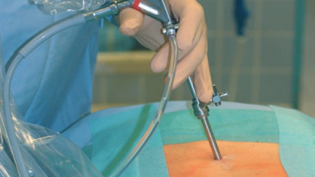
In the decision to perform the operation from the front or back approach in cervical hernia, factors such as the location of the cervical hernia and the experience of the surgeon are important factors.For the approach from the front, the right side of the neck is usually used.After a 4 cm horizontal incision, the subcutaneous tissue, the superficial muscle layer just below it is crossed and the jugular vein is advanced between the neck muscles until the jugular vein is seen.In order to reach the spine, the jugular vein is taken to the outer side, the food and windpipe to the inner side with special retractors and the anterior part of the cervical spine is reached.Again at this stage, the nerve that provides the movement of the right vocal cord and the size of the pupil and the nerve junction that regulates the eyelid movement are worked closely.All these important anatomical structures are carefully protected at every stage.If the surgery will take a long time, to loosen the brackets that we call the retractor that allows us to see the operation site every half hour; It prevents the tissues from being under long-term pressure and ensures normal blood supply.In order to determine the intervertebrae to be operated, x-rays are taken during the operation and the operation site is confirmed.Then the retractors are placed. After this stage of the operation, microdiscectomy is performed under the microscope.In this approach, the drained disc material is replaced with prostheses or bone to fix the two adjacent vertebrae.Then, the operation distance is checked with x-ray for the last 1 time and after the bleeding control, the incision site is closed so that no stitches need to be taken and the operation is terminated.Posterior surgery in cervical hernia is more limited.If the hernia is not in the midline and is in the mouth of the canal where the nerve root from the spinal cord enters to leave the spinal canal, then a posterior approach may be recommended.
As a result, although it is necessary to reach the place to be operated among the very important anatomical structures in the cervical hernia surgery performed by frontal intervention, the complication rates are very low as a result of the careful protection of these structures.When surgery is decided, your physician, who is a neurosurgeon, will tell you in more detail about all kinds of possibilities.The experience we have gained over the years is that the patient's belief in the physician with the physician-patient cooperation always affects the surgical process very positively.TRUST YOUR DOCTOR.
Sağlıkla kalın…